
 Specification
 Support
Super Green I (~Sybr Green) (10000X) Nucleic Acid Stains
Product Description:
Super Green I (ideal substitute for Sybr Green I) Nucleic Acid Stains are produced by Dr.Chimin Du, are a kind of novel generation of fluorescent nucleicacid gel stains designed to replace the highly toxic ethidium bromide (EtBr). Sybr Green I is nontoxic and more sensitive than EtBr. Gels can be visualized under UV or Visible Light.
Features of Sybre Green Nucleic Acid Stains
1. Safety: Sybr Green is nontoxic and noncarcinogenic.
2. Ultra-sensitivity: It allows the visualization of as little as 20pg dsDNA, around 5-10 times more sensitive than EtBr under UV and 8-20 times more sensitive than EtBr in Visible Light.
3. Convenience: NO need to rinse or wash gels. Add stain before load samples. Visualize gels under UV or Visible Light to avoid UV damageon DNA/RNA.
4. Wide range: suitable for agrose gel or PAGE.
5. No impact for the next experiments such as RT, PCR, enzyme digestion, and ligation.
6. Strong signal and no background
Directions for the Agrose Gels stained by Sybr Green
Protocol 1: Pre-cast Protocol (Add dye in the gel)
1.1 Prepare molten agarose gel solution using your standard protocol.
1.2 Add 1~3µl Sybr Green Nucleic Acid Stain per 50ml gel when the gel cool down to 50℃and mix thoroughly.
1.3 Cast the gel and allow it to solidify. Any leftover gel solution may be stored and reheated later for additional gel casting.
1.4 Load samples and run the gels using your standard protocol.
1.5 DNA stained with SYBR Green I stain can be readily visualized using a UV or blue-light sources (emit at 450, 473, 488, or 532 nm). Image the stained gel with the transilluminator and photograph the gel using.
Protocol 2: Post-staining Protocol (Stain Nucleic Acid after electrophoresis by adding dye in the gel stain solution)
2.1 Make gels: Do not add any nucleic acid stain when make gels.
2.2 Run gels as usual according to your standard protocol.
2.3 Prepare Sybr Green Nucleic Acid Staining solution: Dilute Sybr Green Nucleic Acid Stain with TAE or TBE (TBE (89 mM Tris base, 89 mM boric acid, 1 mM EDTA, pH 8) and TAE (40 mM Trisacetate,1 mM EDTA, pH 8) on ratio 1:10000. Stain gels in the dark for 10-30min. Staining time depends on gel concentration and thickness. PAGE can be stained directly on the glass. Let staining solution cover PAGE gels for 30min. please use glassware to store staining solution or silicified glassware because stain will absorb on the glass.
2.4 Visualize gels a UV or blue-light sources.
2.5 Exact molecular weight can be measured by this method but the dyes are used much more in this way.
Protocol 3: Stain nucleic acid before electrophoresis (add dye in the loading buffer)
3.1. Prepare working solution: Dilute 10 µl Sybr Green Stain with 1ml running buffer TBE or TAE. This solution is stable up to one month at 4℃
3.2. Make gels: based on the routine method. Do not add any DNA/RNA stain in the gel.
3.3. Stain Nucleic Acid: Add 1µl Sybr Green I Nucleic Acid Stain working solution to 10µ mixture of sample and loading buffer, let it stay at RT for 3-5min for stain binding to nucleic acid completely. Normally, 1µL working solution is enough for one sample loading, and 1ml Sybr Green I Nucleic Acid Stain is enough to load 10,000 samples.
3.4. Stain markers: Mix 5µL Marker and 1µL Sybr Green I Nucleic Acid Stain working solution thoroughly, let it stay at RT for 5min to let Sybr Green I Stain and DNA/RNA binding completely.
3.5. Load samples and run gels.
3.6. Visualize gels in UV or Visible Light to avoid UV damage on DNA/RNA.
*The big DNA fragments (>2Kb) will move slowly when bind to the stain. So please stain in DNA after electrophoresis or add stain in gels to measure molecular weight exactly.
Notes :
1. Do not run gels over 2 hrs. Or smeared bands appeared because Sybr Green I Stain will dissociate from DNA/RNA.
2. Sybr Green I Stain can dissociate from nucleic acids in ethanol.
3. Please stain nucleic acid in the gel or after electrophoresis to check the exact molecular weight of fragments when compared with molecular weight markers.
4. Please use EP tubes and other plastic wares in Sybr Green I Stain storage, dilution, and staining. Sybr Green I Stain can bind to glassware.
NOTE: Always wear lab coats, gloves and goggles when working with our products although they are low-risk chemicals for R&D only.
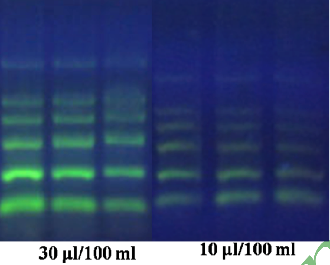
Super Green I 核酸染料不同使用方法电泳图谱:


EB Super Green I DNA Stain EB Super Green I
Fig.1胶染法Fig.1 DNA marker 2000 10, 5, 2.5 per lane Fig.2胶染+点染 Fig.2 DNA Marker 2000 incubate at RT for 3~5min


EB Super Green I DNA Stain EB Super Green II RNA Stain
Fig3. 点染法Fig.3 Volume ration of Dye/DNA marker 2000 Fig.4 Incubate at RT for 3~5min then load 0.5,1,1ug per lane
=1:10 incubate at RT for 3~5min then load 5,2,1uL per lane. Fig4. Sybr Green II点染(RNA)
| Name | Super Green I Nucleic Acid Stains (~Sybr Green) | ||
|---|---|---|---|
| CAT# | 001-100uL | CAS# | N/A |
| Storage# | 4°C干燥避光保存 | Shelf Life# | 12个月 |
| Ex(nm)# | 497 | Em(nm)# | 525 |
| MW# | N/A | Solvent# | DMSO |
| Name | Super Green I Nucleic Acid Stains (~Sybr Green) |
|---|---|
| CAT# | 001-100uL |
| CAS# | N/A |
| Storage# | 4°C干燥避光保存 |
| Shelf Life# | 12个月 |
| Ex(nm)# | 497 |
| Em(nm)# | 525 |
| MW# | N/A |
| Solvent# | DMSO |